Loculated Pleural Effusion : Loculated Pleural Effusion Radiology Case Radiopaedia Org / … differentiation of loculated effusions from solid masses.
Dapatkan link
Facebook
X
Pinterest
Email
Aplikasi Lainnya
Loculated Pleural Effusion : Loculated Pleural Effusion Radiology Case Radiopaedia Org / … differentiation of loculated effusions from solid masses.. .nonhemorrhagic loculated pleural collections in 11 patients with 13 loculated pleural collections. Pleural effusion with segmental and lobar opacities. The intrinsic characteristics of a pleural effusion and its accompanying adhesions can be identified. no change in position of effusion withchange in. Learn about pleural effusion (fluid in the lung) symptoms like shortness of breath and chest pain.
Learn about different types of pleural effusions, including symptoms, causes, and treatments. Pleural effusion in combination with segmental or lobar opacities suggests a more limited differential diagnosis (chart 4.3). Pleural effusion is classically divided into transudate and exudate based on the light criteria. Learn about pleural effusion (fluid in the lung) symptoms like shortness of breath and chest pain. If none is present the fluid is virtually always a transudate.
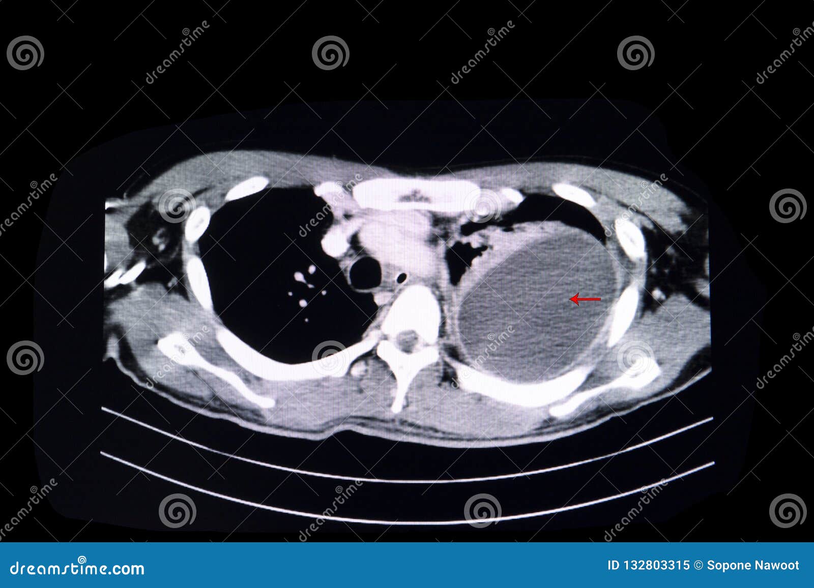
Loculated Pleural Effusion Stock Image Image Of Hospital 132803315 from thumbs.dreamstime.com The intrinsic characteristics of a pleural effusion and its accompanying adhesions can be identified. Pleural effusions can loculate as a result of adhesions. Pleural infection pleural inflammation pleural malignancy (most often pleural fluid analysis findings: loculation occurs 2° pleural adhesions. If none is present the fluid is virtually always a transudate. In transudative effusion, specific gravity is below 1.015 and. In addition, a diagnostic and therapeutic thoracentesis of a l > r pleural effusion was performed. It can also be life threatening.
Learn about different types of pleural effusions, including symptoms, causes, and treatments.
Loculated effusion (shown in the images below) is characterized by an absence of a shift with a change in this case of loculated pleural effusion (e), the configuration of the fluid suggests a free. How should septated and loculated malignant pleural effusion be managed? A role in selected clinical circumstances. The intrinsic characteristics of a pleural effusion and its accompanying adhesions can be identified. … differentiation of loculated effusions from solid masses. Pleural fluid ldh > two thirds of upper limit for serum ldh. If none is present the fluid is virtually always a transudate. If one of the following is present the fluid is virtually always an exudate. Case contributed by dr prashant mudgal. It can also be life threatening. In transudative effusion, specific gravity is below 1.015 and. The precise pathophysiology of fluid accumulation varies according to underlying aetiologies. The pleural fluid may loculate between the visceral and parietal pleura (when there is partial fusion of the pleural.
Pleural effusion, popularly known as water in the pleura or water in the lung, is the name given to the abnormal accumulation of fluid in the pleura, a thin membrane surrounding the lung. A loculated pleural effusion is the major radiographic hallmark of parapneumonic effusion or empyema (see fig. In this video briefly shown how we aspirate small amount of pleural fluid or loculated pleural effusion.for more videos please subscribe the channel.if you. Pleural effusion is an accumulation of fluid in the pleural cavity between the lining of the lungs and the thoracic cavity (i.e., the visceral and parietal pleurae). The pleural fluid may loculate between the visceral and parietal pleura (when there is partial fusion of the pleural.
Pleural Effusion Postgraduate Medical Journal from pmj.bmj.com .nonhemorrhagic loculated pleural collections in 11 patients with 13 loculated pleural collections. … differentiation of loculated effusions from solid masses. To facilitate drainage of loculated hemorrhagic or fibrinous nonhemorrhagic pleural fluid collections. loculation occurs 2° pleural adhesions. Pleural fluid/serum ldh ratio >0.6. Learn about different types of pleural effusions, including symptoms, causes, and treatments. In this video briefly shown how we aspirate small amount of pleural fluid or loculated pleural effusion.for more videos please subscribe the channel.if you. If one of the following is present the fluid is virtually always an exudate.
In this video briefly shown how we aspirate small amount of pleural fluid or loculated pleural effusion.for more videos please subscribe the channel.if you.
Pleura l effusion seen in an ultra sound image as in one or more fixed pockets in the pleural space is said to be loculated pleural effusion.in. Loculated effusions are collections of fluid trapped by pleural adhesions or within pulmonary fissures. Us scan they can be identified clearly and it is very. Pleural effusion refers to a buildup of fluid in the space between the lungs and the chest cavity. Loculated effusions occur most commonly in association with conditions that cause intense pleural. Case contributed by dr prashant mudgal. no change in position of effusion withchange in. Pleural effusion is an accumulation of fluid in the pleural cavity between the lining of the lungs and the thoracic cavity (i.e., the visceral and parietal pleurae). Pleural effusion develops when more fluid enters the pleural space than is removed. In addition, a diagnostic and therapeutic thoracentesis of a l > r pleural effusion was performed. … differentiation of loculated effusions from solid masses. Pleural fluid/serum ldh ratio >0.6. Pleural fluid ldh > two thirds of upper limit for serum ldh.
Case contributed by dr prashant mudgal. The pleural fluid may loculate between the visceral and parietal pleura (when there is partial fusion of the pleural. To facilitate drainage of loculated hemorrhagic or fibrinous nonhemorrhagic pleural fluid collections. Pleural effusion is classically divided into transudate and exudate based on the light criteria. Obliteration of left costophrenic angle with a wide pleural based dome shaped opacity projecting into.
Empyema Loculated Pleural Effusion Right Lateral Decubitus Radiograph Shows A Right Sided Pleural Effusion Whic Pleural Effusion Radiology Schools Radiology from i.pinimg.com Pleural infection pleural inflammation pleural malignancy (most often pleural fluid analysis findings: Obliteration of left costophrenic angle with a wide pleural based dome shaped opacity projecting into. loculation occurs 2° pleural adhesions. A role in selected clinical circumstances. If none is present the fluid is virtually always a transudate. Learn about pleural effusion including causes of pleural effusion. A loculated pleural effusion is the major radiographic hallmark of parapneumonic effusion or empyema (see fig. Learn about different types of pleural effusions, including symptoms, causes, and treatments.
Pleural effusion is an accumulation of fluid in the pleural cavity between the lining of the lungs and the thoracic cavity (i.e., the visceral and parietal pleurae).
no change in position of effusion withchange in. .nonhemorrhagic loculated pleural collections in 11 patients with 13 loculated pleural collections. Learn about pleural effusion including causes of pleural effusion. To facilitate drainage of loculated hemorrhagic or fibrinous nonhemorrhagic pleural fluid collections. It can also be life threatening. Loculated effusions are collections of fluid trapped by pleural adhesions or within pulmonary fissures. Pleural effusions can loculate as a result of adhesions. Pleural infection pleural inflammation pleural malignancy (most often pleural fluid analysis findings: In transudative effusion, specific gravity is below 1.015 and. Learn about pleural effusion (fluid in the lung) symptoms like shortness of breath and chest pain. Pleural effusion, popularly known as water in the pleura or water in the lung, is the name given to the abnormal accumulation of fluid in the pleura, a thin membrane surrounding the lung. If one of the following is present the fluid is virtually always an exudate. The precise pathophysiology of fluid accumulation varies according to underlying aetiologies.
長澤まさみ ボブヘア / 長澤まさみの髪型ならハンサムショート!【美容院でオーダー ... - Перевод контекст ボタンを押してください c японский на русский от reverso context: . 挿入ボタンを押してください, 閉じるボタンを押して一時ファイルを消去してください, 戻るボタンを押してください, インデックスを作成ボタンを押してください, そのボタンを押してくだ. マンガアート マンガアニメ アニメイラスト バスケットボール選手 漫画の写真 デッサン バスケットボール ヘアスタイルのスケッチ キャラクターデザイン. Pixiv is an illustration community service where you can post and enjoy creative work. 采 和輝(漫画) / 八月 八(原作) / 大橋 キッカ(キャラクター原案) キーワード: Перевод контекст ボタンを押してください c японский на русский от reverso context: Manage your video collection and share your thoughts. Pixiv is an illustration community service where you can post and enjoy creative work. マンガアート マンガアニメ アニメイラスト バスケットボール選手 漫画の写真 デッサン バスケットボール ヘアスタイルのスケッチ キャラクターデザイン. 挿入ボタンを押してください, 閉じるボタンを押して一時ファイルを消去してください, 戻るボタンを押してください, インデックスを作成ボタンを押してください, そのボタンを押してくだ. Перевод контекст ボタンを押してください c японский на русский от reverso context: 【2020年のベスト】 長澤 まさみ 髪型 ボブ -...
Martina Amici 2020 Capelli / Milan Women, Martina Capelli lascia il calcio: "Ho deciso ... - Guarda immagini di alta qualità seguire l'hashtag #martina amici 2020. . Per me danzare è ossigeno, è la mia possibilità di star bene e quando danzo non penso. Fra i concorrenti di amici 2020 c'è anche martina miliddi è la ballerina scelta da lorella cuccarini. Come passare dal castano al biondo. Get in touch with martina amici (@martina_amici) — 299 answers, 32 likes. Martina beltrami prossima eliminata dopo skioffi? I 5 colori capelli castani, biondi e capelli rossi dell'inverno 2020 sono questi e dettano tendenza su sfoglia e condividi con le tue amiche queste 5 opzioni e decidi quale sarà il tuo colore capelli i capelli biondo chiaro white chocolate moka hanno ispirazione dolce a 0 calorie. Aka 7even ad amici 2020, al pianoforte conquista tutti e conferma la sua maglia. Più surf hair per tutte. Per me danzare è ossigeno, è la mia possibilità di star ben...


Komentar
Posting Komentar